Overview
Contact
What Is Ankle Cartilage Restoration?
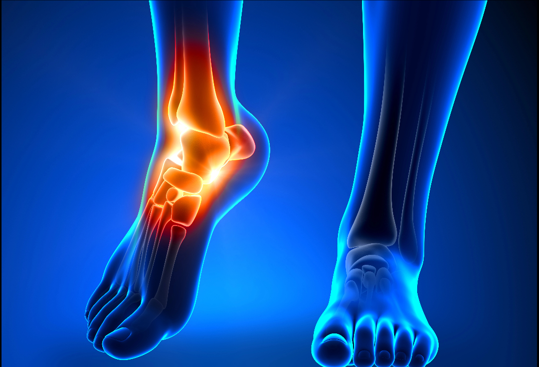
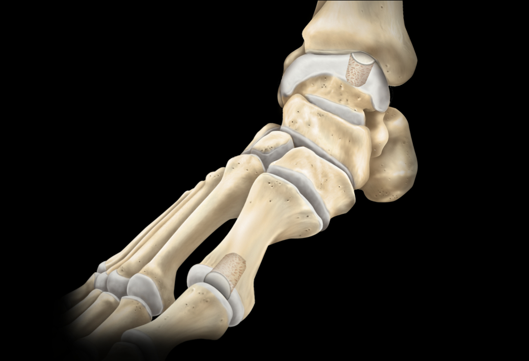
The ankle joint’s articular cartilage is a thin (1-2 mm), resilient hyaline layer covering the talar dome, tibial plafond, and fibular malleolus, facilitating smooth dorsiflexion/plantarflexion (up to 70 degrees total) and subtle inversion/eversion while absorbing compressive forces up to 5 times body weight during gait. Composed of chondrocytes in a matrix of type II collagen, aggrecan, and water, it is avascular and depends on synovial fluid for nourishment. The talus, with its convex dome, bears the majority of load; subchondral bone provides support, while zones include superficial (for shear), transitional (compression), and deep (anchoring). Surrounding ligaments (e.g., ATFL, deltoid) and tendons (e.g., Achilles, peroneals) enhance stability, but the ankle’s high congruency limits intrinsic repair capacity post-injury.
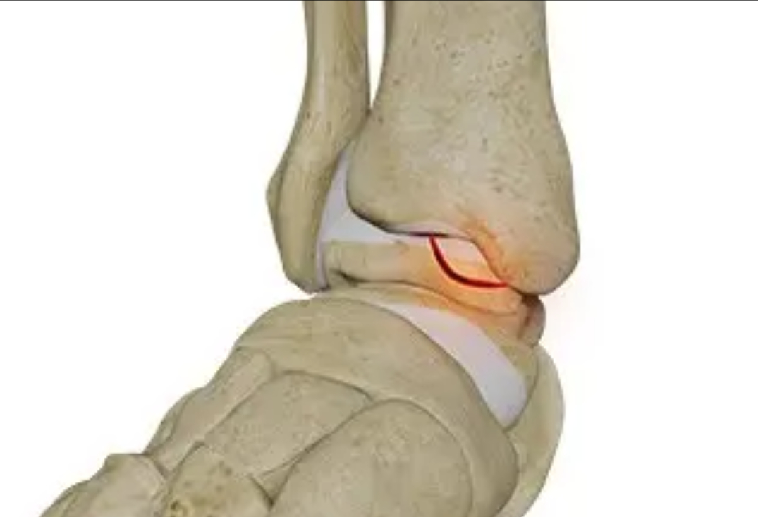
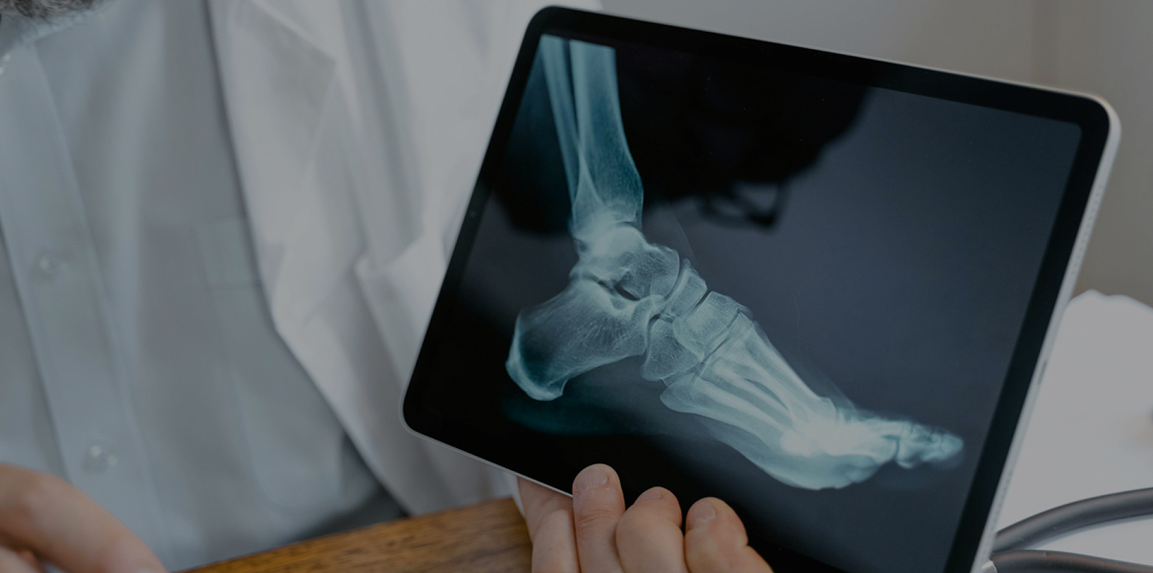
Ankle cartilage defects, often on the talus (osteochondral lesions of the talus or OLTs), arise from trauma (e.g., inversion sprains causing impaction), osteochondritis dissecans (OCD, vascular insult in youth), or repetitive microtrauma (e.g., in soccer or ballet). Graded by Berndt and Harty or ICRS: stage I (subchondral compression), II (partial detachment), III (complete detachment without displacement), IV (displaced fragment). Symptoms include deep ankle pain, swelling, catching/locking, and instability; untreated, defects lead to subchondral cysts, loose bodies, progressive osteoarthritis, and functional decline (e.g., limited dorsiflexion). Medial talar dome lesions (from inversion-plantarflexion) are common; MRI or CT arthrography confirms size (<1.5 cm ideal for some techniques), depth, and cystic changes. Associated instability or alignment issues exacerbate progression.
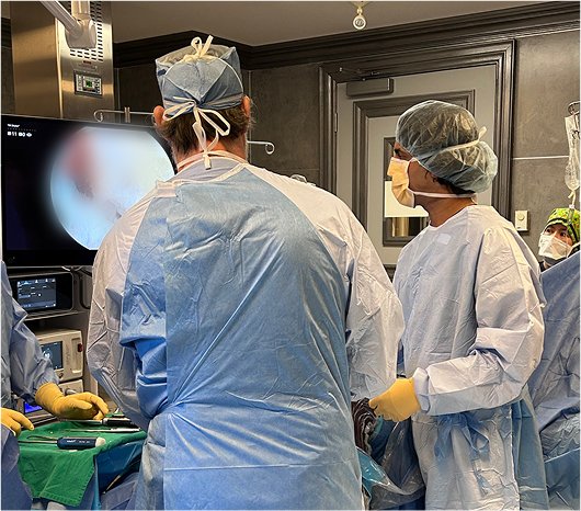
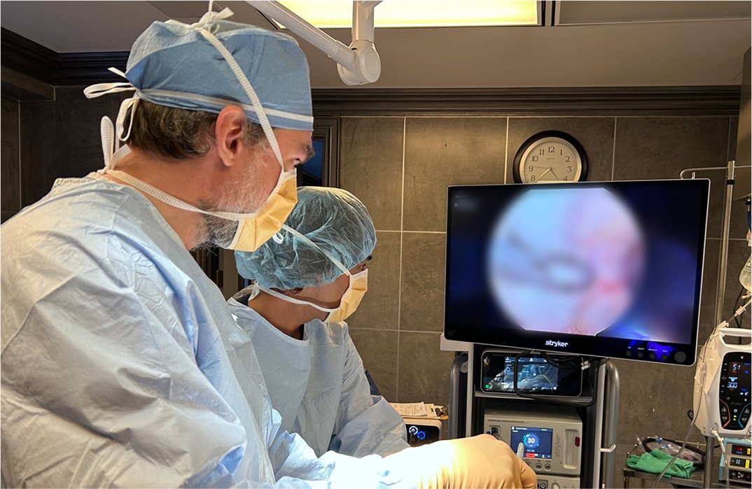
At OV Surgical, we elevate ankle cartilage restoration in Canada with advanced arthroscopic and mini-open techniques that minimize downtime and maximize results, often incorporating orthobiological augmentation with platelet-rich plasma (PRP) or bone marrow aspirate concentrate (BMAC). These orthobiologics, harvested autologously and concentrated for growth factors (e.g., IGF-1, BMPs), are injected or applied to stimulate chondrogenesis, reduce inflammation, and improve integration—enhancing outcomes by 15-30% across methods via MSC recruitment and matrix production. Procedures begin with diagnostic arthroscopy (anteromedial/anterolateral portals) to assess the lesion, debride unstable cartilage to vertical shoulders, and address comorbidities (e.g., synovitis, loose bodies). Defect sizing guides technique; surgery lasts 45-120 minutes.
For full-thickness defects (1-3 cm²), AMIC enhances marrow stimulation. The lesion is debrided, and subchondral bone is microfractured (awls/drills 2-3 mm apart, 4 mm deep) to release MSCs and form a superclot. A collagen matrix (e.g., bilayer membrane) is trimmed, placed over the clot, and secured with fibrin glue or sutures to stabilize cell migration and promote fibrocartilage ingrowth. Orthobiologics like PRP or BMAC are injected beneath the matrix to augment clot quality and accelerate hyaline-like repair.
Suited for contained OLTs (0.5-2 cm²), BioCartilage uses micronized dehydrated allograft cartilage matrix mixed with PRP/BMAC (1:0.8 ratio) for a putty-like fill. Post-debridement and microfracture, the mixture is packed into the defect, leveled flush, and sealed with fibrin glue to prevent washout. This provides a scaffold for host cell infiltration, yielding superior tissue quality vs. microfracture alone; orthobiologics enhance vascularization and chondrocyte viability for better durability.
For smaller defects (<1.5 cm², often cystic), OATS harvests 6-10 mm cylindrical plugs from low-load autologous sites (e.g., knee trochlea or ipsilateral talar non-articular area) using coring tools. The recipient site is reamed to depth, and plugs are press-fit in mosaic fashion for immediate hyaline coverage and bony integration. Orthobiologics (PRP/BMAC) are applied at interfaces to reduce donor morbidity and promote osseous healing, minimizing graft subsidence.
For larger/cystic defects (>1.5 cm² or failed priors), fresh/frozen allografts (talar dome segments) are size-matched via imaging. Through mini-arthrotomy (anterolateral extension if needed), the defect is templated and excised; the allograft is shaped and fixed with press-fit, screws, or pins for structural replacement. Orthobiologics soak the graft or fill voids to boost chondrocyte survival (70-90% viability) and mitigate rejection, improving long-term incorporation.
Recovery
Post-procedure, our evidence-based protocols integrate cryotherapy, neuromuscular electrical stimulation (NMES), and progressive physiotherapy to accelerate healing and restore function, tailored to defect size (small <1.5 cm² vs. larger) and location (weight-bearing talar dome areas require stricter protection). In the immediate protection phase (0-8 weeks), patients are non-weight-bearing with crutches and a boot to minimize shear on the repair site, while allowing passive range of motion (PROM) exercises under therapist guidance to prevent adhesions—starting with continuous passive motion (CPM) machine from 0-30 degrees dorsiflexion/plantarflexion, progressing to 60 degrees by week 2, alongside early isometrics (e.g., toe curls, ankle pumps) to prevent atrophy and maintain circulation. Pain management includes ice, elevation, and anti-inflammatories to control swelling; orthobiologics like PRP or BMAC, applied intraoperatively, can shorten this phase by enhancing tissue viability and reducing inflammation.
From weeks 8-12 (active-assisted phase), crutches are weaned to full weight-bearing, and active-assisted range of motion (AAROM) begins with gentle stretches or aquatic therapy, progressing to full passive ROM while avoiding extreme positions like deep plantarflexion to protect integration. Submaximal isometrics for peroneals, tibialis anterior, and gastrocnemius rebuild neuromuscular control. By weeks 12-16 (active strengthening phase), full weight-bearing continues to be targeted, with resistance exercises like theraband inversion/eversion, calf raises, and balance drills on stable surfaces (2-3 sets of 10-15 reps), emphasizing eccentric control for functional stability. Advanced phases (16+ weeks) incorporate proprioceptive training on wobble boards, agility ladders, plyometrics (e.g., double-leg hops progressing to single-leg), and sport-specific drills (e.g., cutting maneuvers for athletes). Most patients achieve full ROM by 6-8 weeks and strength milestones by 3-4 months, with sports clearance in 6-12 months, verified through tests like ankle strength assessments (>90% symmetry), single-leg balance, hop distances, and in some instances imaging (MRI/CT for integration). Orthobiologics accelerate overall timelines by reducing pain and promoting faster healing. We emphasize psychological readiness, addressing fear of re-injury through gradual exposure.
Benefits
With success rates of 75-85%, ankle cartilage restoration at OV Surgical halts progression, delays osteoarthritis, and allows you to get back to sports and/or other aspects of life that are important to you. Our patients report enhanced ankle function, confidence, and quality of life, backed by meticulous follow-up and data-driven protocols.
FAQ
Contact