Overview
Contact
What Is Ankle Arthroscopy and Stabilization for Instability?
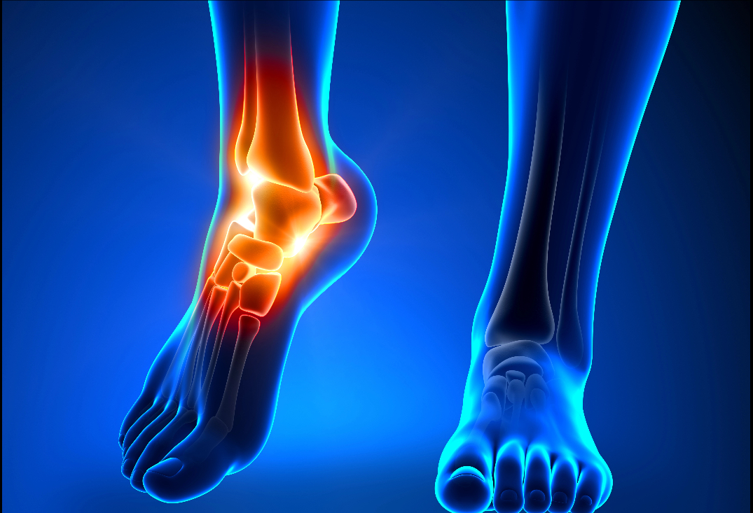
The ankle joint (talocrural) is a hinged synovial articulation between the distal tibia (plafond), fibula (lateral malleolus), and talus dome, enabling dorsiflexion/plantarflexion (20-50 degrees) and subtle inversion/eversion for terrain adaptation. Stability derives from bony congruence (talus wider anteriorly for mortise fit), the joint capsule, and ligaments: medial deltoid (superficial/deep layers resisting eversion) and lateral complex (anterior talofibular ligament or ATFL for inversion restraint in plantarflexion, calcaneofibular ligament or CFL in neutral, posterior talofibular ligament or PTFL in dorsiflexion). The distal tibiofibular syndesmosis (anterior/posterior inferior tibiofibular ligaments, interosseous membrane) maintains fibular position. Articular cartilage covers surfaces for low-friction motion, supported by surrounding tendons (e.g., Achilles posteriorly, peroneals laterally) for dynamic control.
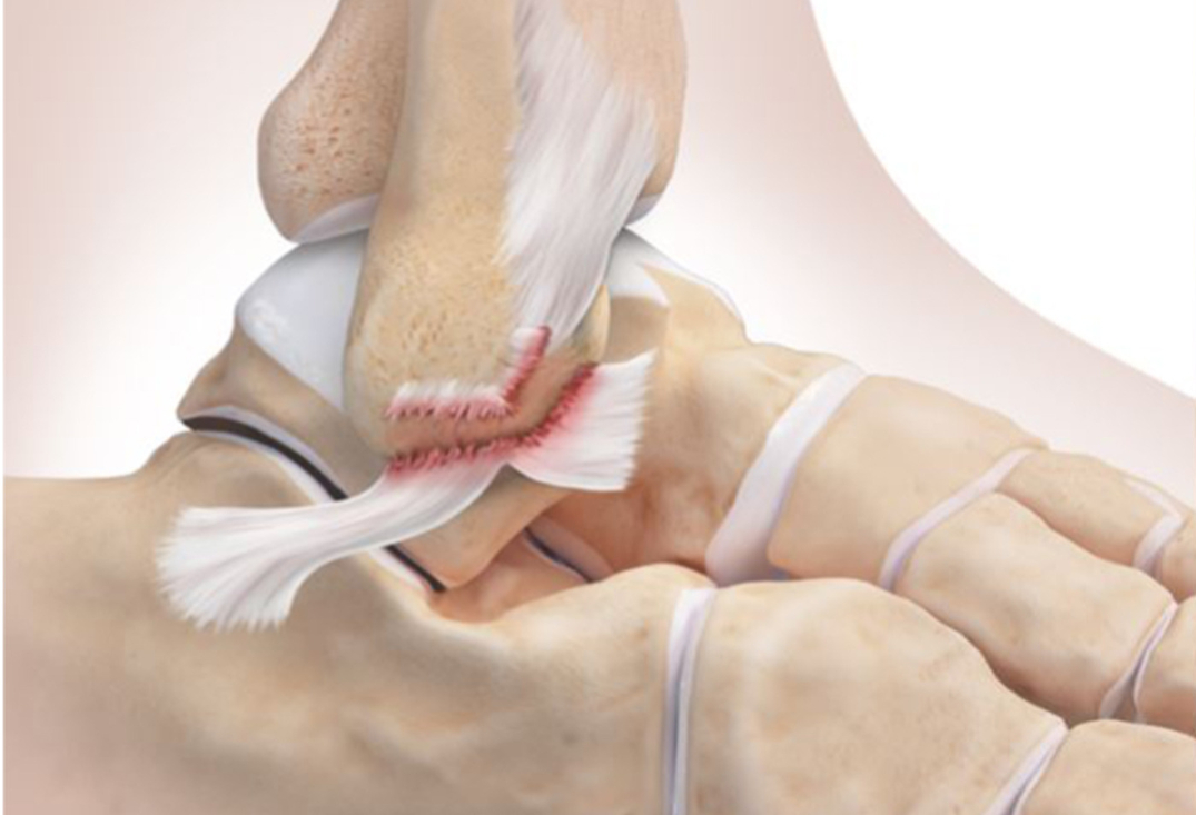
Chronic ankle instability often follows acute lateral sprains (85% of ankle injuries), where inversion forces tear the ATFL (most common, 70% isolated), CFL (20-30% combined), or rarely PTFL. Repetitive microtrauma or incomplete healing leads to ligament laxity, proprioceptive deficits, and mechanical instability, exacerbated by peroneal weakness, hindfoot varus, or syndesmotic injury. Symptoms include recurrent “giving way,” pain on uneven ground, swelling, and positive anterior drawer or talar tilt tests; untreated, it progresses to osteoarthritis from abnormal loading, cartilage wear, and secondary impingement (e.g., anterolateral synovitis or osteophytes). Functional instability may involve neuromuscular issues without anatomic laxity. MRI or stress radiographs confirm ligament damage, loose bodies, or OCD lesions.
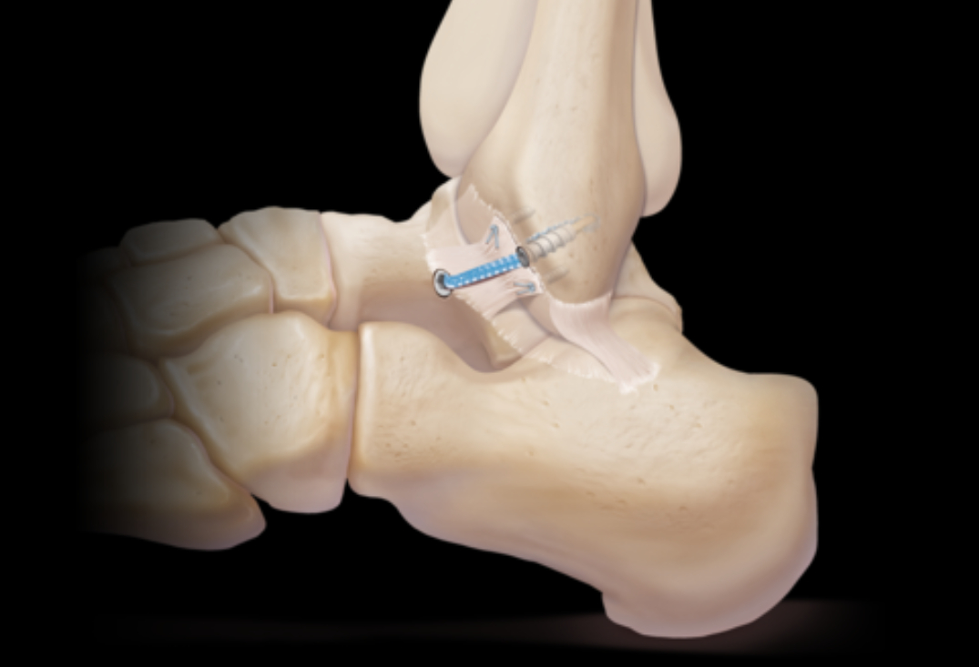
At OV Surgical, we advance ankle arthroscopy and stabilization in Canada with minimally invasive techniques that minimize downtime and maximize results. Performed under general or regional anesthesia with fluoroscopic guidance, the procedure combines diagnostic/therapeutic arthroscopy with ligament reconstruction. Through two to three small incisions (anteromedial, anterolateral portals; posterolateral if needed), an arthroscope visualizes the joint, with fluid distention for space (7-10 mm). Steps include synovectomy/debridement of scarred tissue or impingement (e.g., Bassett’s lesion anterolaterally), removal of loose bodies/osteophytes using shavers or graspers, and assessment of cartilage/syndesmosis. For stabilization, we use the modified Broström-Gould procedure: a curved incision over the lateral malleolus exposes the ATFL/CFL remnants, which are shortened and reattached to the fibula using suture anchors (e.g., 2.9 mm bioabsorbable) in a pants-over-vest fashion for double-layer reinforcement. The inferior extensor retinaculum is mobilized and sutured over the repair for augmentation, enhancing eversion restraint without restricting motion. In severe cases or failed priors, anatomic reconstruction employs autograft (e.g., semitendinosus) tunneled through fibular/talar bone channels. Surgery lasts 60-90 minutes, with closure using absorbable sutures and immediate splinting; orthobiologicals like PRP may be injected for anti-inflammatory and healing effects.
Recovery
Post-procedure, our evidence-based protocols integrate cryotherapy, neuromuscular electrical stimulation (NMES), and progressive physiotherapy to accelerate healing and restore function, tailored to ligament involvement. In the immediate protection phase (0-2 weeks), non-weight-bearing with crutches and a posterior splint or boot to immobilize at neutral, focusing on elevation for swelling control and gentle toe/hip exercises to prevent atrophy. Pain management includes ice and anti-inflammatories.
From weeks 2-6 (early mobilization phase), transition to a walking boot with partial weight-bearing, starting passive range of motion (PROM) under guidance—dorsiflexion to 10 degrees, plantarflexion to 20 degrees—and isometrics for peroneals, tibialis anterior, and gastrocnemius. Stationary biking (no resistance) begins week 4 to promote circulation. By weeks 6-12 (strengthening phase), full weight-bearing in normal shoes, with active range of motion (AROM) progressing to full arcs and resistance exercises like theraband eversion/inversion, calf raises, and balance on stable surfaces (2-3 sets of 10-15 reps). Proprioceptive training on wobble boards rebuilds neuromuscular control while avoiding high-impact. Advanced phases (12+ weeks) incorporate agility drills (e.g., ladder runs), plyometrics (e.g., single-leg hops), and sport-specific simulations (e.g., cutting for soccer players). Most patients achieve full motion by 6-8 weeks and strength milestones by 3 months, with sports clearance in 4-6 months, verified through rigorous testing like single-leg balance (>90% symmetry), star excursion balance tests, and talar tilt exams (<5 degrees side-to-side). We empha
Benefits
With success rates of 85-95% for Broström-Gould, private ankle arthroscopy and stabilization at OV Surgical restores lateral stability, prevents re-sprain, and allows you to get back to sports and/or other aspects of life that are important to you. Our patients report enhanced ankle function, confidence, and quality of life, backed by meticulous follow-up and data-driven protocols.
FAQ
Contact